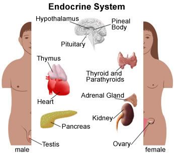
Monday, April 28, 2008

Respiratory System Models

A few more fun A&P links:

lists of all organ systems at innner body.com
interactive A&P learning with getbodysmart.com

Photobucket
body model 3
Terms to recognize and identify
Trachea-
Lungs-
Pulmonary arteries and veins-
Diaphragm-
Larynx-
Carina-
Hyoid bone-
Bronchi-
Primary Bronchus-
Secondary Bronchus-

Photobucket

Photobucket


Photobucket

Photobucket

open heart model

Reminder: When it comes to arteries and veins of the heart
Arteries lead away from the heart (including pulmonary artery leading to lungs)
Veins lead toward the heart (including pulmonary veins with oxygenated blood)

Endocrine System- the hormonal system perhaps :)

Endocrine System:

Photobucket

For a quick explanation of the endocrine system check out the University of Montana web page. It also explains the effects of alcohol on the endocrine system which would be pertinent for many college students and more than one of my friends :) The web site really isn't very specific... and it leaves out the thymus... the alcohol reference reminded me of college students in general, so I thought to include it.
For a more all encompassing and in depth explanation check out Lake Michigan College their web site is very comprehensive and is quickly becoming my favorite online reference for this class.

pituitary gland
-Pituitary gland: small marble-sized gland located in the brain directly below the hypothalamus. The gland has two parts, the anterior and posterior; it excretes many hormones as we should all know from lecture.
thyroid gland-Two lobes, located in front of the windpipe just below the voice box.
Produces two hormones thyroxine and triiodothyronine: thyroid hormones in general increase the metabolism of almost all body tissues.
parathyroid gland-Four pea sized bodies located behind the thyroid gland that produce PTH. Our lab instructor stated it just looks like "larger bumps" on the under-side (posterior) of the thyroid. SO IF IT IS PINNED it will be the pin posterior to the thyroid.
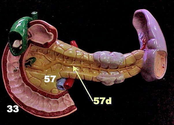
pancreas- Located in abdomen behind (deep to) the stomach. It releases pancreatic secretions into the pancreatic duct which joins with bile and dumps into the duodenum. The secretion's release is controlled by the hepatopancreatic sphincter, also called the Sphincter of Oddi. Some people have an accessory pancreatic duct which has it's own entry into the duodenum (see associated digestive organs post below.)
Thymus- Located in mediastinum and lies on pericardium. In the cadaver it is merely a flap over the heart region because it shrinks in size as we age... and our cadaver looks OLD.
Adrenal glands- Are located on superior border of kidneys. As we remember from histology, they are comprised of three zones of Adrenal Cortex (zona glomerulosa, zona fasiculata & zona reticularis).
Photobucket

endocrine system

Oral Cavity including Teeth and Salivary Glands

A short list to know for lab:

Parotid Gland- The largest gland behind the tongue in the back of the throat
Sublingual Gland- below the tongue
Submandibular Gland- below the mandible

tonsils model

Oral Cavity:
Terms to know and identify:
Lingual Frenulum- connects tongue and gingiva. Lift your tongue and it's the flap of skin below
Labia- from the Latin word for "lips" ... because it means lips.
Labial Frenulum- flap of skin above top teeth connecting labia and gingiva
Gingiva- The tissue of the upper and lower jaws that surrounds the base of the teeth. Also called gums, an infection/inflammation here is called gingivitis.

Remember to know the dental formula.

dental formula

teeth

Digestive Tract- The result is a load of...

Photobucket

Digestive Tract:
Esophagus
Stomach
Fundus
- small superior portion
Body
Pylorus
- bottom 1/3 of stomach
Pyloric Sphincter- inferior sphincter of stomach into Duodenum of small intestine
Cardiac Sphincter- superior sphincter of stomach behind heart
Rugae-large irregular folds of stomach inner lining
Greater Curvature
Lesser Curvature
Greater Omentum
Lesser Omentum


stomach regions

Small Intestine
Duodenum
Hepatopancreatic sphinter (Sphincter of Oddi
)- from hepatopancreatic duct
Plica circulares
- All segments of the small intestine have plicae circularis which are folds of submucosa that increase surface area. Villi are finger-like projections of the lamina propria, upon and between plicae circulares
Jejunum- Often found in umbilical area of cadavers
Ileum- Lower portion of small intestine ending in Ileocecal valve
Ileocecal Valve
- Leeds to cecum

Cartoon Colon
colon labeled

Cecum- the balloon like structure at the base of the Colon
Appendix- extends off cecum

The Colon is divided into 5 sections
ascending colon
transverse colon
descending colon

sigmoid colon- "S" shaped
rectum- the end of the line

Teniae Coli- longitudinal layer of smooth muscle comprised of 3 bands
(tenia coli singular)
Haustra- pocket like sacs
(Haustrum singular)
Right Colic flexure
Left Colic flexure


Photobucket
colon model
body model 2
GI tract

Anatomy of Associated Digestive Organs

The digestive tract has a number of associated organs we should be able to identify on models or cadavers... Of course the cadavers are too star-style to have their pictures taken unless it's paparazzi style, and I just can't do that to a sweet old (dead) man. SO here's hoping you can pick them out in the flesh too. a Montana University has been nice enough to put cadaver organ pics up

For a great overview of the entire digestive system photos et al check out Lake Michigan College's web site Many photos here in this lab section have been from their collection. There are simple descriptions and tutorials. main A&P page at Lake Michigan Comm College Cruise the site in between my-spacing or face-booking all your buddies... Check out the interactive colon; it's loads of fun!
Here's a liver picture you can find with interactive labels on the afore mentioned web site:
liver 4

Associated Organs
What to know for the test:
Liver
Quadrate Lobe
Caudate Lobe
Left Lobe
Right Lobe
Falciform Ligament
- divides right and left liver portions
Gall Bladder
Hepatic Ducts
Cystic Duct
- duct from common bile duct to gallbladder to store excess bile
Common Hepatic duct- bile leaves the liver toward gall bladder and duodenum
Common Bile Duct
- bile toward pancreatic duct and duodenum
Hepatic Artery
Hepatic Portal Vein
Hepatic Vein
Pancreas
Pancreatic Duct
Accessory pancreatic duct
Duodenal Ampulla


Pictures from lab models
liver model

liver model 2

Bile duct drawing
bile duct