Tuesday, June 3, 2008

Male Gross Anatomy.. no that's what HE said

Do you need a quick review of Histology slides without journeying to an open lab??? Check out this web site... you can literally ZOOM IN on the slides online. Sweet! Thanks to
University of Iowa

As always, my all time favorite SIU med web site their histology is comprehensive respiratory to reproductive with great explanations in only a few sentences.


Terms to Know: *** know on cadaver if *** asterisks present Sorry I need pics of the models... be careful however if googling such titles... Haha
Scrotum***- skin and fascia
testes***-
Septa
Tunica Albuginea***- white cord most inner
Tunica Vaginalis***- double layered covering
Epididymis***
Spermatic Cord***- in the cord contains/ holds all
Pampiniform plexus- testes veins
Cremaster muscle- helps regulate temperature with proximity
Ductus Deferens (vas deferens)
Ampulla
Ejaculatory duct
Urethra
Prostatic- through prostate
membranous- below prostate to penis short
Spongy Penile Urethra ***


Penis
Corpus Spongiosum ***- holds urethra in middle
Corpus Cavernosa***-
Glans Penis***-extension of spongiosa
Prepuce*** (foreskin)

Prostate Gland- 1/3 seminal fluid
Seminal Vesicles- 2/3 seminal fluid
Bulbourethral glands- clear viscous pre-ejaculate
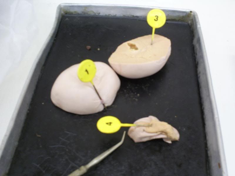
testes
1- Tunica albuginea
3- Septum
4- Epididymis
Female Anatomy Plenty to grow in...

Ovary
Hilium- vessels enter ovary
Corpus Luteum
Graafian Follicle
Oviduct-
Fimbriae- fingers connected to fallopian tubes gather oocyte
Infundibulum- next to fimbriae
Ampulla- large region where fallopian tubes turn
Isthmus- connect fillopian tubes to uterus


Uterus:
Endometrium- inner portion which is shed
Myoetrium- thich muscular wall
perimetrium- outside wall
Round ligament- connects uterus to abdominal wall

Cervix- extention of uterus to vagina
Cervical canal
broad ligament
uterus
vagina
mons pubis
labia majora (s. labium majus)
labia minora (s. labium minus)
clitoris
Breast
nipple
areola
mammary duct- produces milk
lactiferous sinus- milk stored
Colostrum- yellowish made first and second day after egg implantation


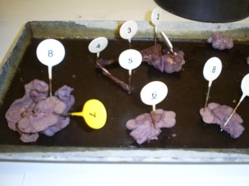
Actual Class dissections labeled:
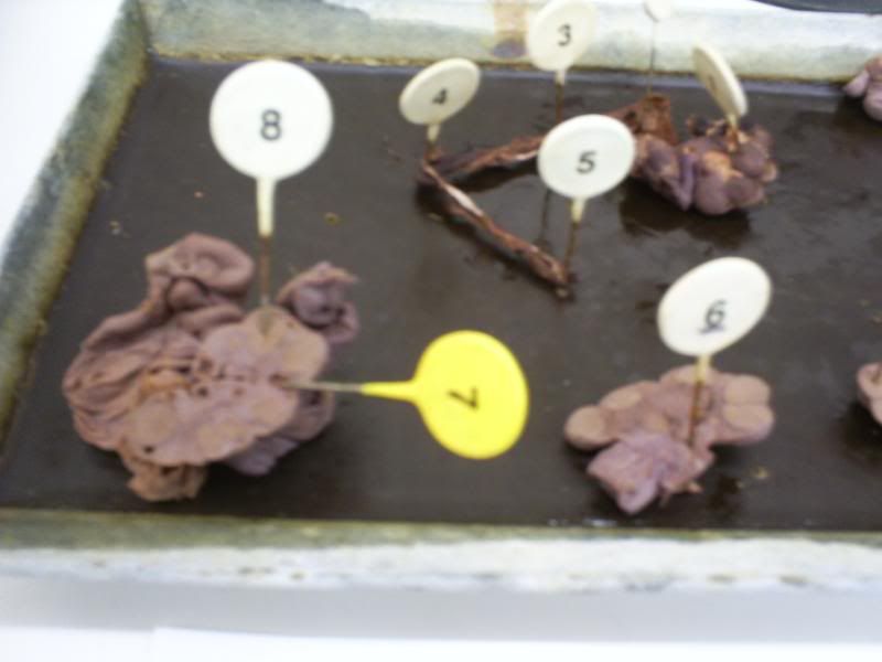
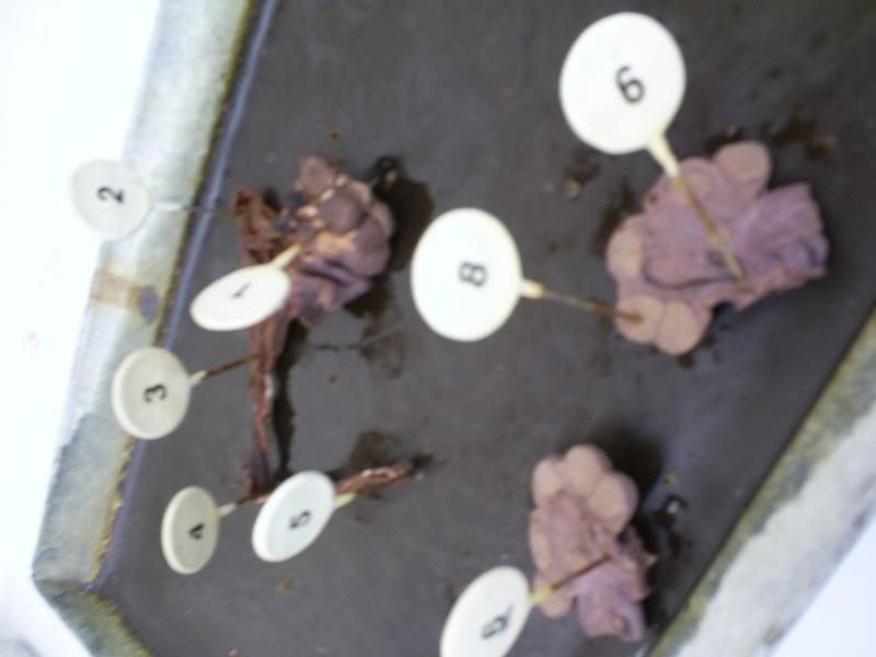
Ovary Gross Anat

White Pins-
1-Ovary
(Oviduct)
2- fimbriae
3- infundibulum
4- Ampulla
5- Isthmus
6- Hilum
7- Graafian Follicle
8- Corpus Luteum


Ovary gross Anat
Ovary Gross Anat

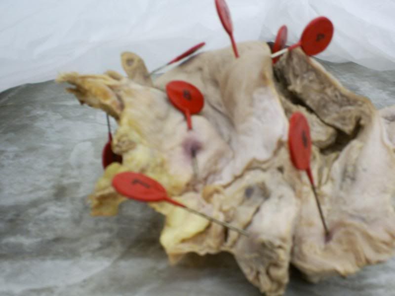
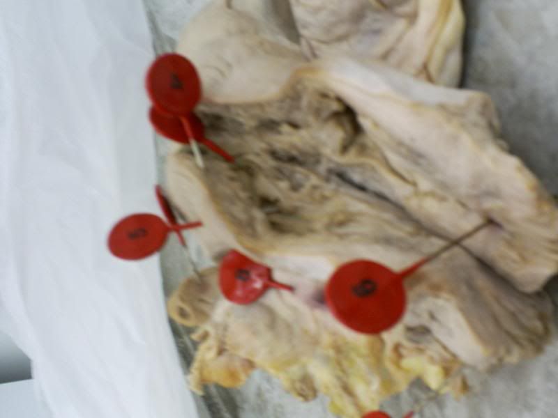
Uterus Pinned:
Uterus

Red Pins-
1- Ovary
2- Oviduct
Uterus
3- endometrium
4- Myometrium
5- perimetrium
6- Cervical Canal
7- Cervix
8- Broad Ligament

Photobucket

Monday, June 2, 2008

Testes and Male hisotology- guys are like SO easy!

Testes:
terms to know:
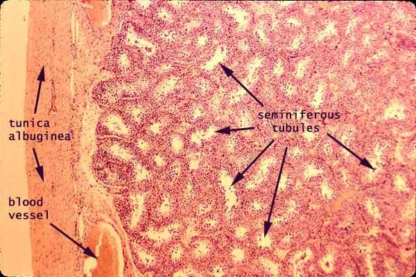
Seminiferous Tubules- tunica albugenia extending into septum
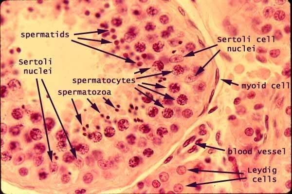
Sertoli cells- cell with dark nucleus clear outside
Spermatogonia- dense to outside less developed
Spermatocytes- less denseley packed
Spermatids- smaller late form with a tail
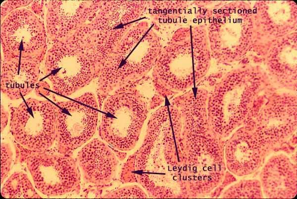
Interstitial cells (Cells of Leydig)- between seminiferous tubules
Tunica Albuginea
Septa

Testes Seminiferous Tubules
Photobucket

testes leydig cells


Epididymis:
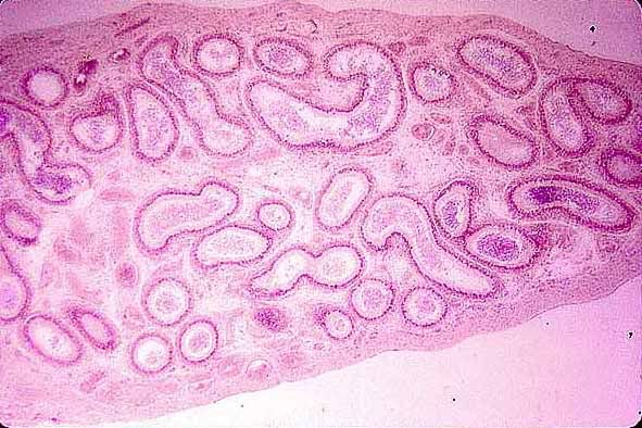
Efferent Ductules- long ducts carry sperm to epididymis- wavy edged lumes with spermatazoa in center
Ductus Epididymis-
Stereocilia- line the epididymis
Spermatozoa
Testes Epididymis


Vas Deferens:
Testes Vas Deference


Penis:
Glands of Littlre
Corpora Cavernosa-
Corpus Spongiosum- contains urethra to center
Urethra
Tunica Albuginea- covers corpora cavernosa


Secretory Glands:
Seminal Vesicle- production 2/3 semen fluid in this highly folded mucosa
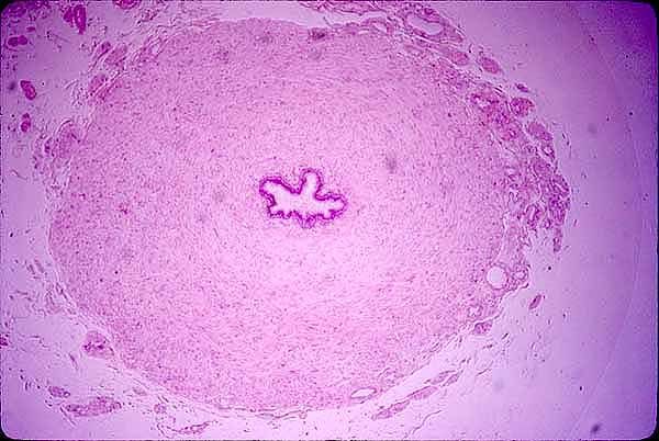
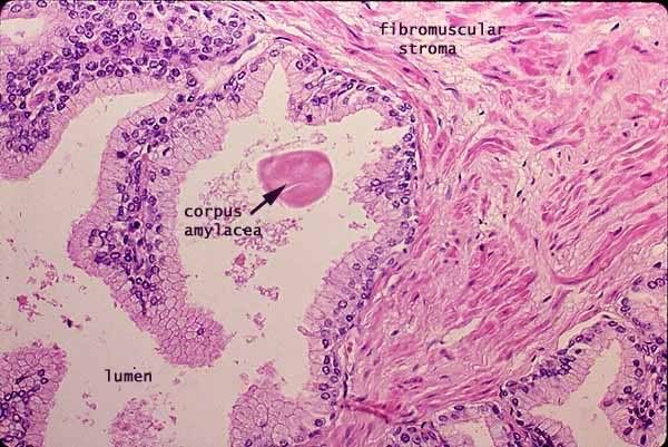
Prostate
Prostatic Concretions- main thing in gland look glossy and pink

Photobucket

Spermatic cord-
Pampiniform plexus- veins near ductus deferens
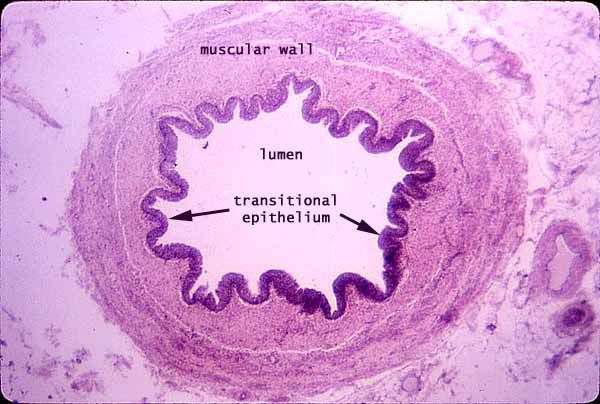
Smooth Muscle-
longitudinal
circular
male urethra

Female Ovary Histology- should it be HERstology?

Ovary terms to know:
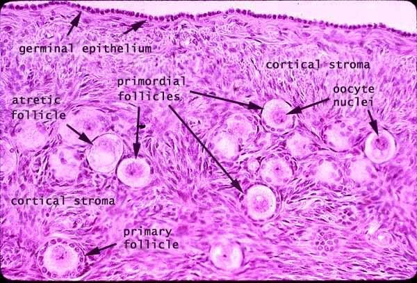
tunica albuginea- between germinal epithelium and cortex
germinal epithelium- outtermost layer
Primordial follicle- earliest stage found before birth in women has very small s squamous surrounding
Primary Follicle- 2nd stage simle cuboidal layer
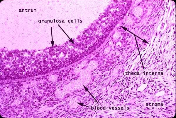
Secondary Follicle- antrum in cell, thecal cells surrounding
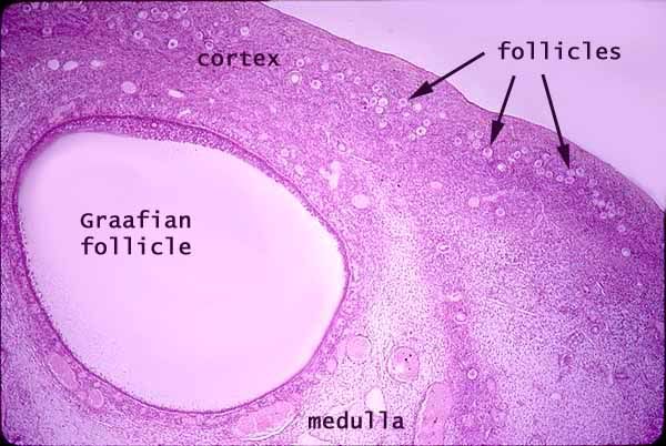
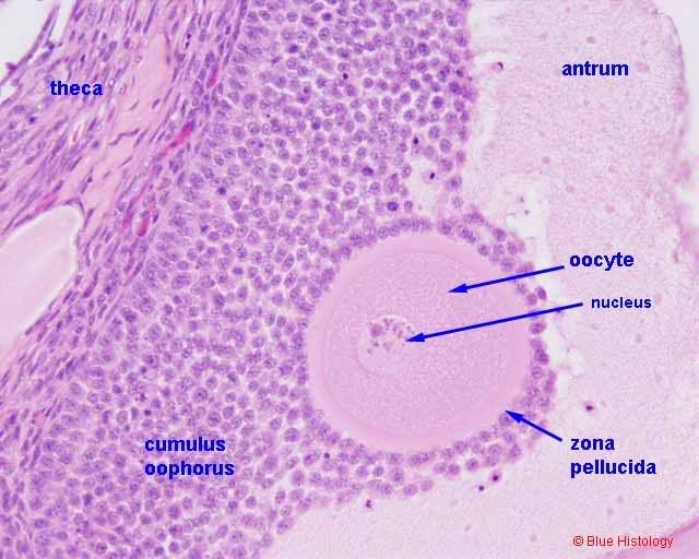
Graafian Follicle- has stand/pedestal called cumulous oophorus
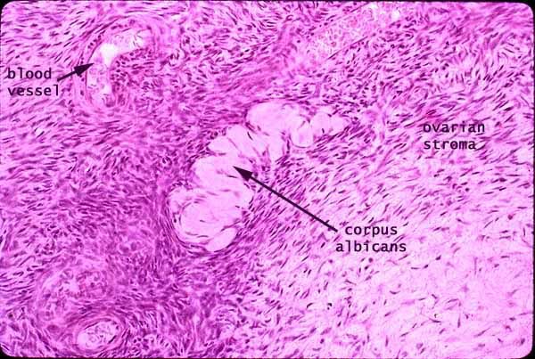
Corpus luteum- empty space after oocyte is expelled produces progesterone if oocyte fertilized and implanted
Antretic Follicle- results if oocyte not implanted
Antrum- open space filled with follicular fluid- fluid signifies 2nd follicle in late stage, the off follicle becming Graafian follicle
Follicular Fluid
Oocyte
Primary oocyte
Secondary oocyte
zoa pellucida- pink ribbon glycoprotein between granulosa cell
corona radiata- protects oocyte by bathing it with estrogen
cumulus oophorus
theca interna- outside follicle darker area
follicular cells

Photobucket

Photobucket

Photobucket

Photobucket
Photobucket

To Womb it May concern Uterine and Menstrual Histo

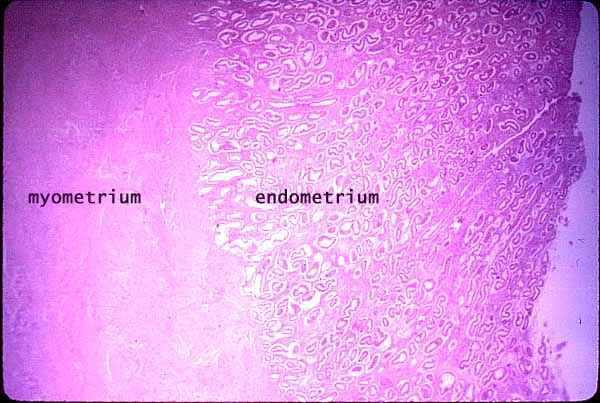
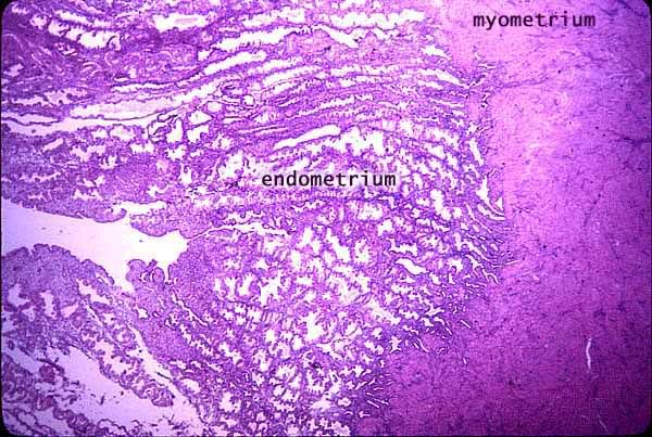
Uterine Histology

Endometrium- functional layer which is expelled monthly
Basal Layer- Layer Not expelled in menses from which spiral veins grow
Functional Layer- contain uterine gland
Myometrium- Muscular layer deep to endometrium
Perimetrium- outer lining of uterus
Endometrial Glands
Venous sinuses- also called lakes form in endometrium during menses and are shed
layers of uterus

uterus


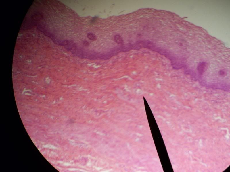
Vagina Histology:
Stratified Squamous epithelium- contains a darker stain than the stratified squamous of anal canal. The Stratification and tight cell junctions prevent against injury as on the skin. Unlike skin these are unkeratinized USUALLY, excessive alcohol and drugs can actually begin keratinization of these cells.
Vascular Submucosa- deep region with blood vessells

Uterus Stratified Squamous

Menstrual Stages of Histology (NOTE: these differ from the physiological/hormonal stages)
Week 1- Menstrual Week superficial layer is torn with veins and arteries present
Week 2- Proliferative week not as thick repairing superficial layer
Week(s) 3-4 Secretory Week thick with endocrine and gland activity, also contains cork screw shape

No slides per TA of menstrual cycle