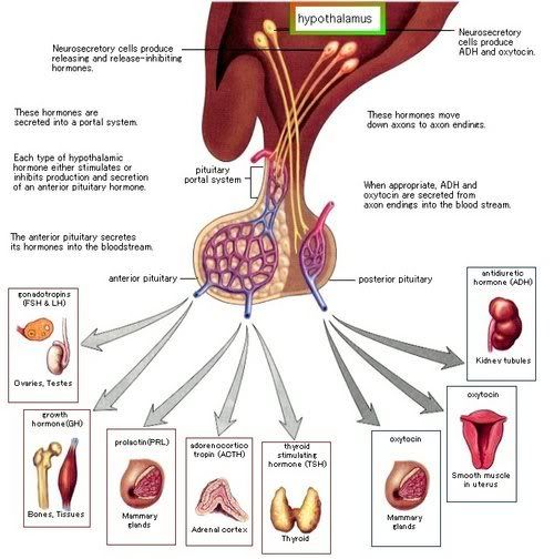
The pituitary, or hypophysis, is really two distinct glands.
1. The anterior pituitary, also called the adenohypophysis.
2. The posterior pituitary, also called the neurohypophysis.
Both parts are attached by the pituitary stalk (also called neural stalk/ infundibulum) to the median eminence at the base of the hypothalamus.
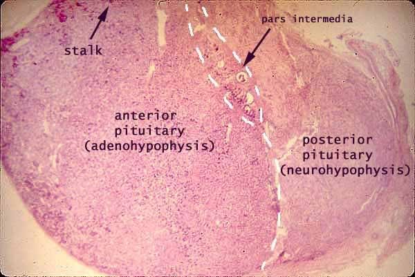
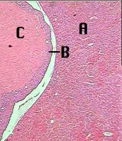
At 40x the pars distalis (A) and the pars intermedia (B) of the adenohypophysis (anterior pituitary) and the pars nervosa (C) of the neurohypophysis (posterior pituitary) can be observed. The pars tuberalis and infundibulum were not a part of this preparation. The pars distalis secretes Growth Hormone (GH), Thyroid-stimulating hormone (TSH), Adrenocorticotrophic hormone (ACTH), Follicle-stimulating hormone (FSH), Lutenizing hormone (LH),and Prolactin. The pars intermedia secretes Melanocyte-stimulating hormone (MSH). The pars nervosa stores ADH (antidiuretic hormone)and Oxytocin which were secreted by the hypothalamus.
Anterior Pituitary:
Terms to know and Recognize:
Pars Tuberalis
Pars Intermedia
Pars Distalis
Chromophobes- "color" phobes, don't stain well, nucleus appears in hollow hole
hypophyseal portal system
tropic hormones- hormone of anterior pituitary
MSH- melanocyte-stimulating hormone
Acidophils- secrete GH and PRL (Growth Hormone and Prolactin)
Basophils- secrete FSH, LH, ACTH, TSH (Follicle stimulating hormone FSH and leutinizing hormone LH are both referred to as gonadotrophs, adrenocoricotropic hormone ACTH, thyrotropic hormone TSH)
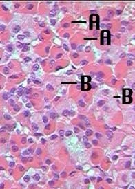
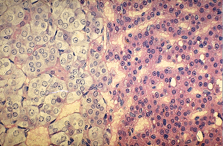
At higher magnifications the dark staining chromophils ( A) and the very light staining chromophobes (B) are easily distinguished.
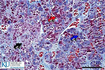
Chromophobes (black arrow), Eosinophiles (red arrow), Basophiles (blue arrow).
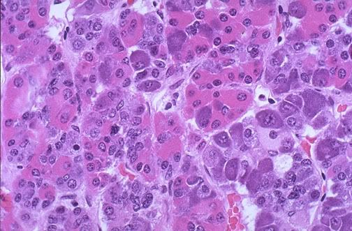
The normal microscopic appearance of the adenohypophysis is shown here. The adenohypophysis contains three major cell types: acidophils, basophils, and chromophobes. The staining is variable, and to properly identify specific hormone secretion, immunohistochemical staining is necessary. A simplistic classification is as follows:
The pink acidophils secrete growth hormone (GH) and prolactin (PRL)
The dark purple basophils secrete corticotrophin (ACTH), thyroid stimulating hormone (TSH), and gonadotrophins follicle stimulating hormone-luteinizing hormone (FSH and LH)
The pale staining chromophobes have few cytoplasmic granules, but may have secretory activity.
The Hypophyseal Portal system and Hormones explained simply
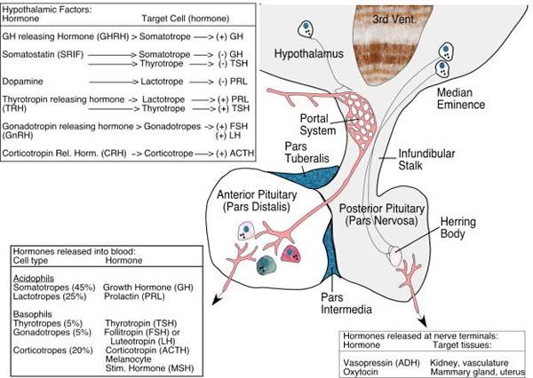
A portal system is a blood vessel circuit with 2 sets of capillaries instead of 1. The first capillary bed (1 capillary plexus) picks up the hypothalamic hormones from the ends of the axons. The second capillary bed (2 capillary plexus) drops off the hypothalamic hormones in the anterior pituitary. Connecting the 2 capillary beds are the hypophyseal portal veins.
Posterior Pituitary
Terms to Know:
Pars nervosa
Hypophyseal Tract- blood vessel portal with only one segment as in posterior pituitary
Pituicytes
Neural stalk- (aka) infundibulum
oxytocin
ADH- antidiuretic hormone
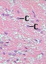
This region of the pituitary is non secretory. Its cells are neuroglial-like pituicytes (C).
Wednesday, April 16, 2008
Endocrine Histology Thyroid and Parathyroid
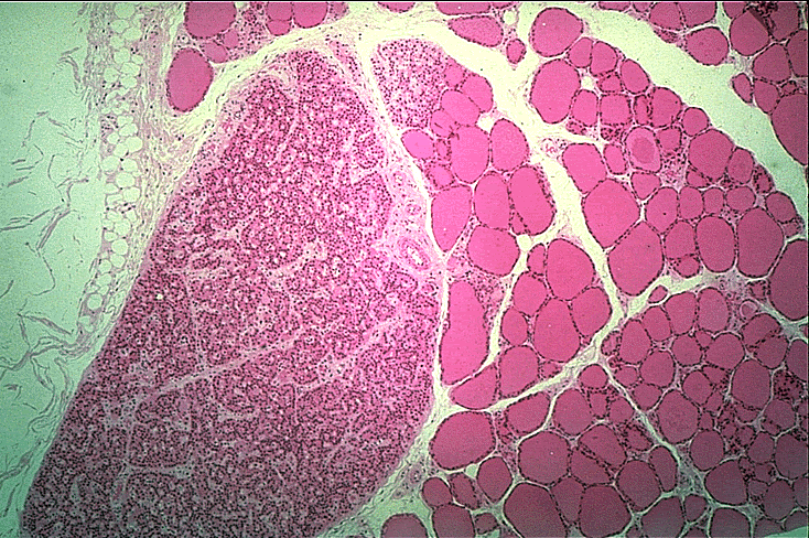
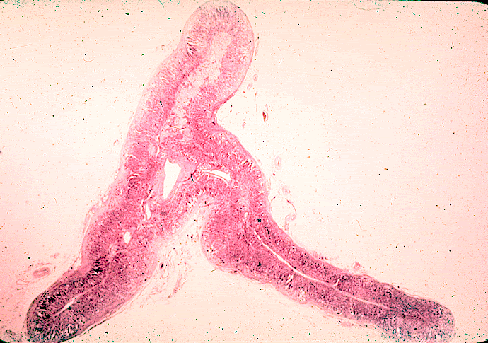
The Thyroid and Parathyroid are located in the same area and can be found in the same slides:
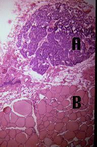
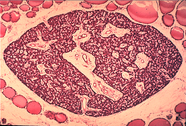
Parathyroid in (A) the egg shaped dark stain in this picture with(B) Thyroid
Thyroid
Terms to know:
Colloid
Follicular Cells- produce hormones T3 and T4
Follicles
Parafollicular Cells (C-cells)- produce Calcitonin
Thyroid Hormone
Sinusoids
Thyroid Follicles
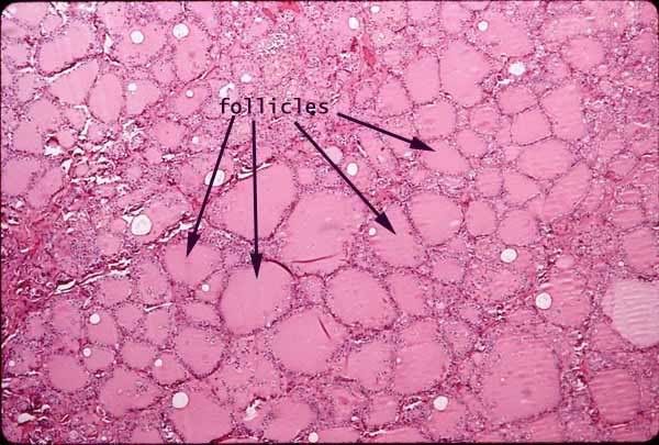
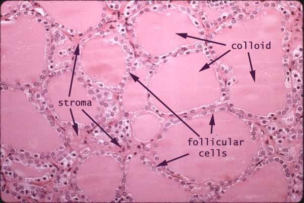
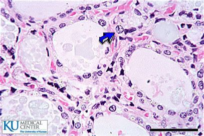
Thyroid follicles and colloid The simple cuboidal epithelium lining the follicles produces the thyroglobulin which is stored in the colloid follicles. Later it is taken back up by these same cells, cleaved, and released as T3 & T4.
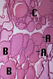
The thyroid gland is composed of many spherical hollow sacs called thyroid follicles. In this tissue section, each follicle (A) appears as an irregular circle of cells. The principal cells, which surround the follicle are simple cuboidal epithelium. These follicles are filled with a colloid (B), which usually stains pink. The principal cells use the thyroglobulin and iodide stored in the colloid to produce the primary thyroid hormones - including thyroxine.
Between these follicles are the parafollicular cells (C) which produce calcitonin.
Parafollicular cells (C Cells) release Calcitonin.
Parathyroid:
Terms to Know:
Chief Cells- secrete parathyroid hormone (PTH)
oxyphil cells
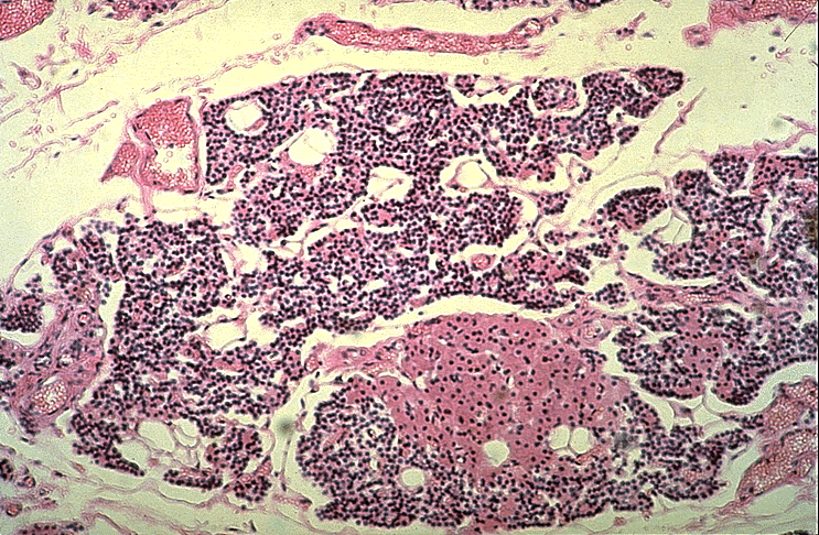
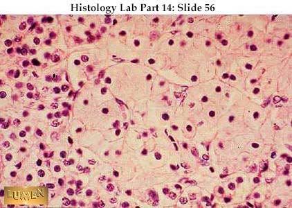
The parathyroid consists chiefly of chief cells (duh!). The chief cells are small cells arranged into curvilinear cords. Parathyroid chief cells secrete parathyroid hormone (PTH)(again, duh!), which stimulates osteoclast activity and thus raises the blood calcium level. PTH thus works antagonistically with calcitonin (from thyroid C cells) to regulate blood calcium.
The parathyroid also contains oxyphil cells (see next photo)
Parathyroid at higher power, showing that the cells are actually tightly packed epithelial cords. The larger, pale pink cells in the middle and to the right are oxyphil cells; they have a smaller, darker nucleus and relatively larger amount of cytoplasm than the majority of cells, which are called chief cells (to the left in the photo). The chie f cells secrete parathormone; the significance of the oxyphil cells is not clear.
For more Thyroid/Parathyroid fun check out Lumen Lab's Slides
Endocrine System Adrenal Gland
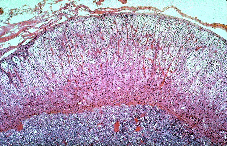
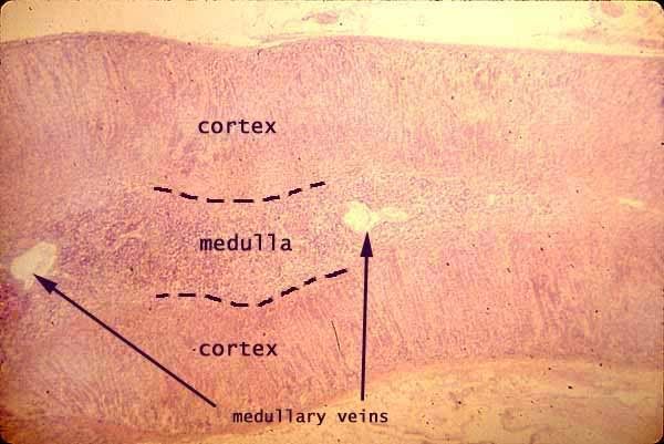
The adrenal Gland is comprised of the capsule, cortex and medulla areas.
The Adrenal Cortex:
Terms to know and recognize:
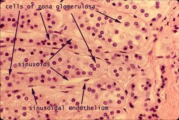
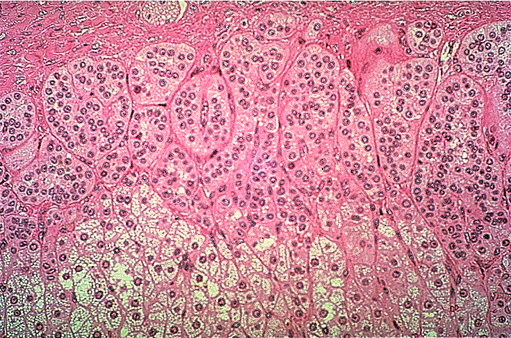
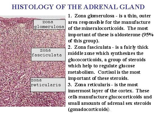
Zona Glomerulosa- produces hormone Aldosterone
Zona Fasciculata- produces hormone Cortisol
Zona Reticularis- produces sex hormones Androgen
Sinusoids
Adrenal Sinusoids
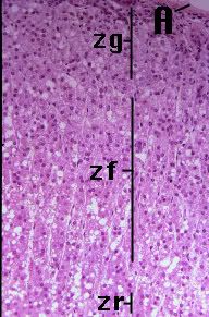
The Zona's
Zona Glomerulosa- Aldosterone
Zona Fasciculata- Cortisol
Zona Reticularis- Androgen
Zona Glomerulosa up close
Zona fasciculata and reticularis
Unmarked all Zona's
Adrenal medulla:
terms to Know:
Chromaffin cells- produce both adrenaline and noradrenaline
Sympathetic ganglia
Medullary Vein- Adrenal blood supply flows from capsular arteries to medullary veins by percolating through cortical sinusoids.
Chromaffin Cells
Medullary vein
A Few pictures to review the adrenal gland
Zona Reticularis and Fasciculata
All zonas
Endocrine Histology Pancreas, Thymus and Pineal Body
Pancreas
Know the following:
Islets of Langerhans
alpha and beta cells
exocrine acinar tissue
glucagon- produced by alpha cells
insulin- produced by beta cells
Full view of Pancreas- Pancreatic islets of Langerhans are small nests of cells, arranged into curvilinear cords, scattered throughout the pancreas.
Islets are usually conspicuously paler (less intensely stained) than the acini (OR Exocrine Acinar Tissue) of the exocrine pancreas, but in any case islets differ markedly from exocrine pancreas in the arrangement of cells (cords rather than acini).
Pancreatic islets contain several endocrine cell types secreting insulin (beta cells), glucagon (alpha cells), somatostatin (delta cells), and pancreatic polypeptide (PP cells). These cells cannot be readily distinguished in routine preparations but may be identified with special stains.
Special Stains for Alpha and Beta Cells (do not need to recognize for test)
Immunoperoxidase staining can help identify the nature of the cells present in the islets of Langerhans. On the right, antibody to insulin has been employed to identify the beta cells. On the left, antibody to glucagon identifies the alpha cells.
Alpha cells secrete glucagon
Beta cells secrete insulin
Islets of Langerhans
Exocrine Acinar Tissue
Thymus:
Terms to learn:
Cortex
Medulla
Thymosin
Lymphoid tissue
Another view of Thymus
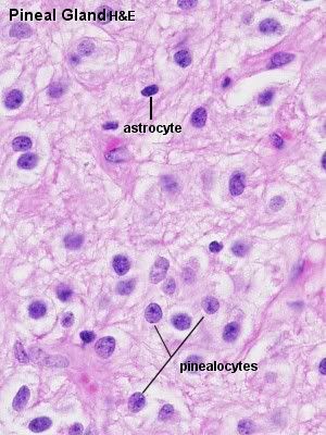
Pineal Body:
Know the following: (it's a short list)
concretions (brain sand)
melatonin
Labels:
Anatomy and physiology,
endocrine,
histology,
pancreas,
pineal body,
thymus
Endocrine Histology Plenty of Histo Ovaries and testes
Ovary Medulla and Cortex
Terms to know:
Estrogens
medulla
Cortex
Progesterone
follicles
inhibin
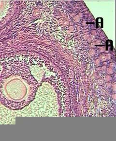
The outer portion of the ovary is called the cortex. The inner portion is called the medulla. There is some ill-defined difference in stromal texture between cortex and medulla, but the main distinction is that the cortex includes all of the ovarian follicles while the medulla contains the larger blood vessels.
Ovarian Follicles
Ovarian Follicles produce estrogen, progesterone and inhibin
Ovarian Follicles
Testes:
Terms to know:
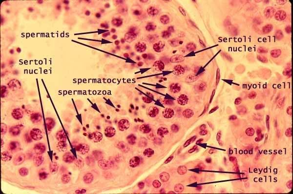
Interstitial Cells (cells of Leydig)- produces testosterone
Sustentacular cells (Sertoli cells)- produces inhibin
Sustentacular Cells, also called Sertoli cells nourish spermatogenic cells
form blood/testis barrier to keep sperm from immune system. Produces Inhibin
Cells of Leydig produce Testosterone
Interstitial cells, also called cells of Leydig
There is a plethora of Histology gathered here for the class lab, most are found on various University websites... Although there are probably a handful, or less students who check this blog (counting myself of course) to give credit where credit is due the slides are gathered from the following web pages:
University Delaware Biology
University of Omaha
Kansas State University
My Favorite Histology Site so far from the school of medicine
SIU School of Medicine
Another great Histology resource College of Medicine U of Illinois great practice and info!
For a quick break down of all organs and hormones in one quick page (histology on links) check outLake Michigan College
One Quick Look at one page of histology Endocrine Histology in a glance
Terms to know:
Estrogens
medulla
Cortex
Progesterone
follicles
inhibin
The outer portion of the ovary is called the cortex. The inner portion is called the medulla. There is some ill-defined difference in stromal texture between cortex and medulla, but the main distinction is that the cortex includes all of the ovarian follicles while the medulla contains the larger blood vessels.
Ovarian Follicles
Ovarian Follicles produce estrogen, progesterone and inhibin
Ovarian Follicles
Testes:
Terms to know:
Interstitial Cells (cells of Leydig)- produces testosterone
Sustentacular cells (Sertoli cells)- produces inhibin
Sustentacular Cells, also called Sertoli cells nourish spermatogenic cells
form blood/testis barrier to keep sperm from immune system. Produces Inhibin
Cells of Leydig produce Testosterone
Interstitial cells, also called cells of Leydig
There is a plethora of Histology gathered here for the class lab, most are found on various University websites... Although there are probably a handful, or less students who check this blog (counting myself of course) to give credit where credit is due the slides are gathered from the following web pages:
University Delaware Biology
University of Omaha
Kansas State University
My Favorite Histology Site so far from the school of medicine
SIU School of Medicine
Another great Histology resource College of Medicine U of Illinois great practice and info!
For a quick break down of all organs and hormones in one quick page (histology on links) check outLake Michigan College
One Quick Look at one page of histology Endocrine Histology in a glance
Labels:
Anatomy and physiology,
histology,
ovary,
ovary histology,
testes
Subscribe to:
Posts (Atom)