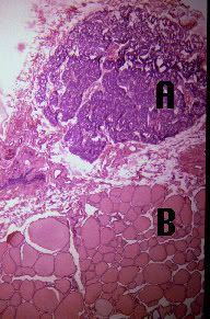
Parathyroid in (A) the egg shaped dark stain in this picture with(B) Thyroid
Thyroid
Terms to know:
Colloid
Follicular Cells- produce hormones T3 and T4
Follicles
Parafollicular Cells (C-cells)- produce Calcitonin
Thyroid Hormone
Sinusoids
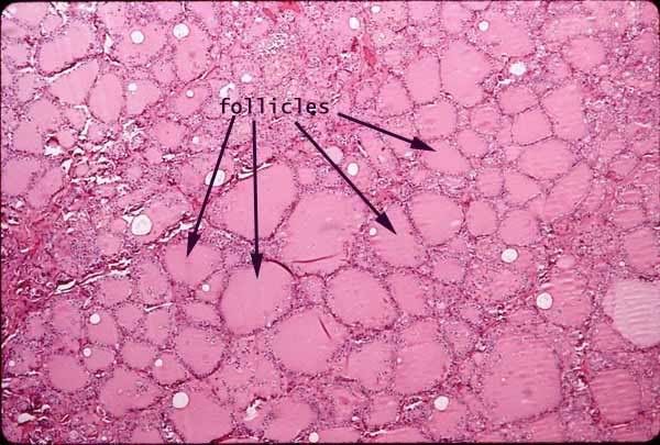
Thyroid Follicles
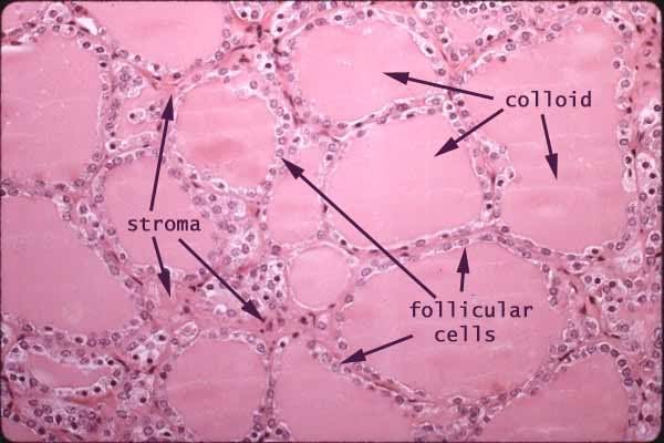
Thyroid follicles and colloid The simple cuboidal epithelium lining the follicles produces the thyroglobulin which is stored in the colloid follicles. Later it is taken back up by these same cells, cleaved, and released as T3 & T4.
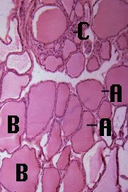 The thyroid gland is composed of many spherical hollow sacs called thyroid follicles. In this tissue section, each follicle (A) appears as an irregular circle of cells. The principal cells, which surround the follicle are simple cuboidal epithelium. These follicles are filled with a colloid (B), which usually stains pink. The principal cells use the thyroglobulin and iodide stored in the colloid to produce the primary thyroid hormones - including thyroxine.
Between these follicles are the parafollicular cells (C) which produce calcitonin.
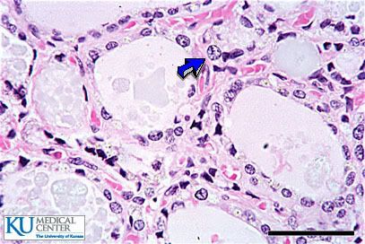
Parafollicular cells (C Cells) release Calcitonin.
Parathyroid:
Terms to Know:
Chief Cells- secrete parathyroid hormone (PTH)
oxyphil cells
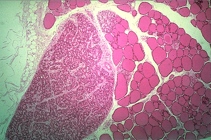
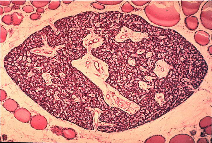
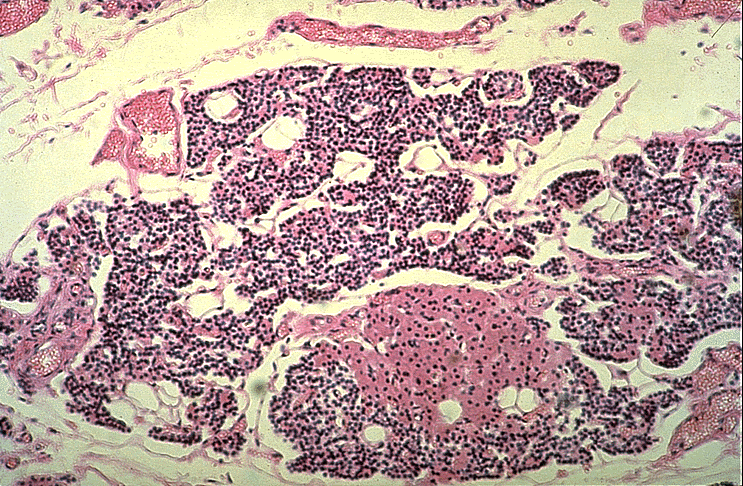
The parathyroid consists chiefly of chief cells (duh!). The chief cells are small cells arranged into curvilinear cords. Parathyroid chief cells secrete parathyroid hormone (PTH)(again, duh!), which stimulates osteoclast activity and thus raises the blood calcium level. PTH thus works antagonistically with calcitonin (from thyroid C cells) to regulate blood calcium.
The parathyroid also contains oxyphil cells (see next photo)
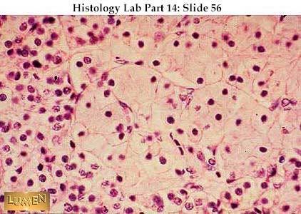
Parathyroid at higher power, showing that the cells are actually tightly packed epithelial cords. The larger, pale pink cells in the middle and to the right are oxyphil cells; they have a smaller, darker nucleus and relatively larger amount of cytoplasm than the majority of cells, which are called chief cells (to the left in the photo). The chie f cells secrete parathormone; the significance of the oxyphil cells is not clear.
For more Thyroid/Parathyroid fun check out Lumen Lab's Slides
3 comments:
Salamu Alaykom!!!
You're total saviour! I have a Histo exam in 3 hours and I discover last night that it's PRACTICAL!!
And Whenever I click on google image I like I get shipped back here!!! The slides really are great!!!!!! Jazaky Allah Khayran " )
Farah Saleh
Din mamma är en gris.
Very Useful,
Shukarran!!
Post a Comment