Monday, May 5, 2008

Stomach, are you hungry?

Stomach what to know:

Rugae
Oblique muscle layer
Gastric pits- areas between inlets
Gastric glands- far lower than pits
Mucous epithelial cells- line top area (unlike goblet cells NOT mixed with regular cells)
Parietal cells- look like fried eggs with a darker center
Zymogenic "chief" cells- all others in gastric gland

*hint* NO villi, NO goblet cells


Photobucket

Most of the bulk of the gastric mucosa is occupied by secretory cells of the gastric glands, primarily parietal cells (P) and chief cells (C), together with lamina propria.

Photobucket

Photobucket

Photobucket
Photobucket

Duodenum thru Colon histo- Oh no... they're all a like

Duodenum:

Brunner's glands
Plicae Circulares

*hint* duodenum contains villi

Photobucket

jejunum:

*hint* long skinny villi and NO Brunner's glands

Photobucket

Ileum:
Peyer's patches

*hint* short squat villi, many goblet cells, lots of lymphatic tissue, few plicae circulares

Photobucket

Photobucket

Peyer's patches clearly shown here
Photobucket

Appendix

*hint* no villi, many goblet cells, fecal concretions, diffuse lymphatic tissue

Photobucket

Lab slides live in your PC- or MAC

KU has the most comprehensive histology easy to cruise through and get all the photos you need. A large number of the histo pics here are from KU... they are labeled as such.

online histo slides

University of Illinois school of Medicine site histo

Try and figure a few out.... or see above posts with hints

salivary intralobular duct

Salivary parotid gland

salivary intralobular duct

Photobucket

Esophageal gland

Monday, April 28, 2008

Respiratory System Models

A few more fun A&P links:

lists of all organ systems at innner body.com
interactive A&P learning with getbodysmart.com

Photobucket
body model 3
Terms to recognize and identify
Trachea-
Lungs-
Pulmonary arteries and veins-
Diaphragm-
Larynx-
Carina-
Hyoid bone-
Bronchi-
Primary Bronchus-
Secondary Bronchus-

Photobucket

Photobucket


Photobucket

Photobucket

open heart model

Reminder: When it comes to arteries and veins of the heart
Arteries lead away from the heart (including pulmonary artery leading to lungs)
Veins lead toward the heart (including pulmonary veins with oxygenated blood)

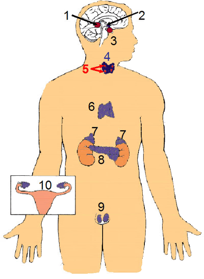
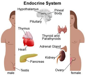
Endocrine System- the hormonal system perhaps :)

Endocrine System:

Photobucket

For a quick explanation of the endocrine system check out the University of Montana web page. It also explains the effects of alcohol on the endocrine system which would be pertinent for many college students and more than one of my friends :) The web site really isn't very specific... and it leaves out the thymus... the alcohol reference reminded me of college students in general, so I thought to include it.
For a more all encompassing and in depth explanation check out Lake Michigan College their web site is very comprehensive and is quickly becoming my favorite online reference for this class.

pituitary gland
-Pituitary gland: small marble-sized gland located in the brain directly below the hypothalamus. The gland has two parts, the anterior and posterior; it excretes many hormones as we should all know from lecture.
thyroid gland-Two lobes, located in front of the windpipe just below the voice box.
Produces two hormones thyroxine and triiodothyronine: thyroid hormones in general increase the metabolism of almost all body tissues.
parathyroid gland-Four pea sized bodies located behind the thyroid gland that produce PTH. Our lab instructor stated it just looks like "larger bumps" on the under-side (posterior) of the thyroid. SO IF IT IS PINNED it will be the pin posterior to the thyroid.
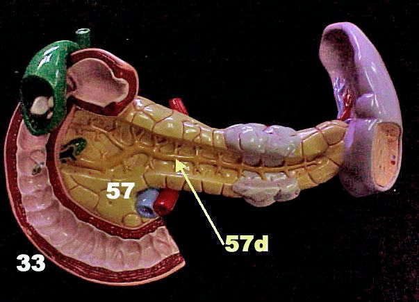
pancreas- Located in abdomen behind (deep to) the stomach. It releases pancreatic secretions into the pancreatic duct which joins with bile and dumps into the duodenum. The secretion's release is controlled by the hepatopancreatic sphincter, also called the Sphincter of Oddi. Some people have an accessory pancreatic duct which has it's own entry into the duodenum (see associated digestive organs post below.)
Thymus- Located in mediastinum and lies on pericardium. In the cadaver it is merely a flap over the heart region because it shrinks in size as we age... and our cadaver looks OLD.
Adrenal glands- Are located on superior border of kidneys. As we remember from histology, they are comprised of three zones of Adrenal Cortex (zona glomerulosa, zona fasiculata & zona reticularis).
Photobucket

endocrine system