Amusing YouTube Tutorial with a simple cat model with veins marked. Less grotesque, although the narrator is bit interesting to listen to.
Wednesday, February 27, 2008


Veins and Arteries- catastrophy
Yes, not savory... Thanks to the street feline who donated his body to science. What can you do? People, it's like Bob Barker says...
Anyways our pics for the week

Arterial System Shown Here
Red Pins:
1- Aortic Arch
2- Right Brachiocephalic Artery
3- Subclavian Artery
4- Common Carotid Artery
5- Axillary Arery
6- Brachial Artery
Venous System
White Pins/Yellow Pins:
1. Superior Vena Cava
2. Inferior Vena Cava
3. Azygos Vein
4. Brachiocephalic Vein
5. External Jugular Vein
6. Inter Jugular Vein
7. Subclavian Vein
8. Axillary Vein
9. Brachial Vein
Another Picture Same View

~~~~~~~~~~~~~~~~~~~
Lower body Venous and Arterial Pins

Arterial System Shown Here
Red Pins:
11. Renal Artery
12. Inferior Mesenteric Artery
13. Iliolumbar Artery
14. External Iliac Artery ** In humans there exist the Lt and Rt common Iliac Arteres
15. Internal Iliac Artery **On cats internal iliac arteries branch off med. Sacral Artery
16. Femoral Artery
Venous System
White Pins/Yellow Pins:
8.Axillary Vein
9.Brachial Vein
10. Renal vein
11. Gonadal vein
12. Common Iliac Vein
13. External Iliac Vein
14. Femoral Vein
15. Great Saphenous Vein
Another Picture same view:

2 More Views: 1 Without pins


Ask the wizard for a heart...
Pictures, slides and links for the heart -- anyone send a few so I can make a collection. Unfortunately never received any pictures from the lab... so on to the next.


Wednesday, February 20, 2008
Histology Blood
Well, it's a bit late, and I get the feeling it's only me using this blog anyways...
None the less, here's the blood histology slides I found online. This week seemed fairly simple:
Formed Elements make up 45% of blood
Plasma makes up 55% of blood
Erythrocytes: Red blood cells

Leukocytes are white blood cells and include the following:
Granulocytes- Neutrophil, Eosinophil, and Basophil appear grainy in slides

Neutrophil: (50-70% of Leukocytes)
multi-lobe neucleus, active phagocyte

Eosinophil: (2-4% of Leukocytes)
bilobed neucleus

Basophil (0.5-1 % of Leukocytes)
"S" shaped neucleus

Agranulocytes: no visible granules clear cyctoplasm
Lymphocyte- spherical neucleus imported for antibody immune response


Monocytes: "U" shaped neucleus, long term-chronic illness 2x's as big as RBC

To remember the greatest percentage in blood/plasma use the achronymn
Never Let Monkeys Eat bananas
neutrophil
lymphocyte
monocyte
eosinophil
basophil
A few more slides

Neutrophil and Eosinophil

Lymphocyte and Neutrophil
None the less, here's the blood histology slides I found online. This week seemed fairly simple:
Formed Elements make up 45% of blood
Plasma makes up 55% of blood
Erythrocytes: Red blood cells

Leukocytes are white blood cells and include the following:
Granulocytes- Neutrophil, Eosinophil, and Basophil appear grainy in slides
Neutrophil: (50-70% of Leukocytes)
multi-lobe neucleus, active phagocyte
Eosinophil: (2-4% of Leukocytes)
bilobed neucleus
Basophil (0.5-1 % of Leukocytes)
"S" shaped neucleus

Agranulocytes: no visible granules clear cyctoplasm
Lymphocyte- spherical neucleus imported for antibody immune response

Monocytes: "U" shaped neucleus, long term-chronic illness 2x's as big as RBC
To remember the greatest percentage in blood/plasma use the achronymn
Never Let Monkeys Eat bananas
neutrophil
lymphocyte
monocyte
eosinophil
basophil
A few more slides

Neutrophil and Eosinophil

Lymphocyte and Neutrophil
Tuesday, February 12, 2008
More pictures from classmates
Click here for Great Crista Ampullaris info online
Great quick animation about balance and the maculae click here
This animation shows the crista ampullaris and the connection with semicircular ducts to maintain balance click here

Cochlea Histology

Sound Reception in the Cochlea

Inner Ear The Cochlea
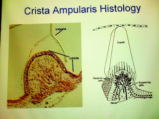
Crista Ampullaris

Crista Ampullaris Histology
Inner Ear Anatomy

Middle Ear Anatomy

The Organ of Corti

Organ of Corti 2

Great quick animation about balance and the maculae click here
This animation shows the crista ampullaris and the connection with semicircular ducts to maintain balance click here

Cochlea Histology

Sound Reception in the Cochlea

Inner Ear The Cochlea

Crista Ampullaris

Crista Ampullaris Histology
Inner Ear Anatomy

Middle Ear Anatomy

The Organ of Corti

Organ of Corti 2

Subscribe to:
Posts (Atom)