Showing posts with label Anatomy and physiology. Show all posts

Wednesday, April 16, 2008

Endocrine Histology Pancreas, Thymus and Pineal Body

Pancreas
Know the following:
Islets of Langerhans
alpha and beta cells
exocrine acinar tissue

glucagon- produced by alpha cells
insulin- produced by beta cells


Full view of Pancreas- Pancreatic islets of Langerhans are small nests of cells, arranged into curvilinear cords, scattered throughout the pancreas.

Islets are usually conspicuously paler (less intensely stained) than the acini (OR Exocrine Acinar Tissue) of the exocrine pancreas, but in any case islets differ markedly from exocrine pancreas in the arrangement of cells (cords rather than acini).

Pancreatic islets
contain several endocrine cell types secreting insulin (beta cells), glucagon (alpha cells), somatostatin (delta cells), and pancreatic polypeptide (PP cells). These cells cannot be readily distinguished in routine preparations but may be identified with special stains.

Special Stains for Alpha and Beta Cells (do not need to recognize for test)


Immunoperoxidase staining can help identify the nature of the cells present in the islets of Langerhans. On the right, antibody to insulin has been employed to identify the beta cells. On the left, antibody to glucagon identifies the alpha cells.
Alpha cells secrete glucagon
Beta cells
secrete insulin


Islets of Langerhans


Exocrine Acinar Tissue

Thymus:
Terms to learn:
Cortex
Medulla
Thymosin
Lymphoid tissue


Another view of Thymus

Pineal Body:
Know the following: (it's a short list)
concretions (brain sand)
melatonin


Pineal body


Endocrine Histology Plenty of Histo Ovaries and testes

Ovary Medulla and Cortex

Terms to know:
Estrogens
medulla
Cortex
Progesterone
follicles
inhibin


ovary medulla and cortex

The outer portion of the ovary is called the cortex. The inner portion is called the medulla. There is some ill-defined difference in stromal texture between cortex and medulla, but the main distinction is that the cortex includes all of the ovarian follicles while the medulla contains the larger blood vessels.


Ovarian Follicles


Ovarian Follicles produce estrogen, progesterone and inhibin


Ovarian Follicles


Testes:
Terms to know:
Interstitial Cells (cells of Leydig)- produces testosterone
Sustentacular cells (Sertoli cells)- produces inhibin


Testes Leydig and Sustentacular Cells

Sustentacular Cells, also called Sertoli cells nourish spermatogenic cells
form blood/testis barrier to keep sperm from immune system. Produces Inhibin


Cells of Leydig produce Testosterone


Interstitial cells, also called cells of Leydig


There is a plethora of Histology gathered here for the class lab, most are found on various University websites... Although there are probably a handful, or less students who check this blog (counting myself of course) to give credit where credit is due the slides are gathered from the following web pages:

University Delaware Biology

University of Omaha

Kansas State University

My Favorite Histology Site so far from the school of medicine
SIU School of Medicine

Another great Histology resource College of Medicine U of Illinois great practice and info!

For a quick break down of all organs and hormones in one quick page (histology on links) check outLake Michigan College

One Quick Look at one page of histology Endocrine Histology in a glance

Wednesday, April 9, 2008

Muscle Tissue Histology

Skeletal Muscle Striated


Skeletal Muscle striated and Multinucleate


Smooth Muscle
Muscle smooth


Smooth Muscle Uninucleate


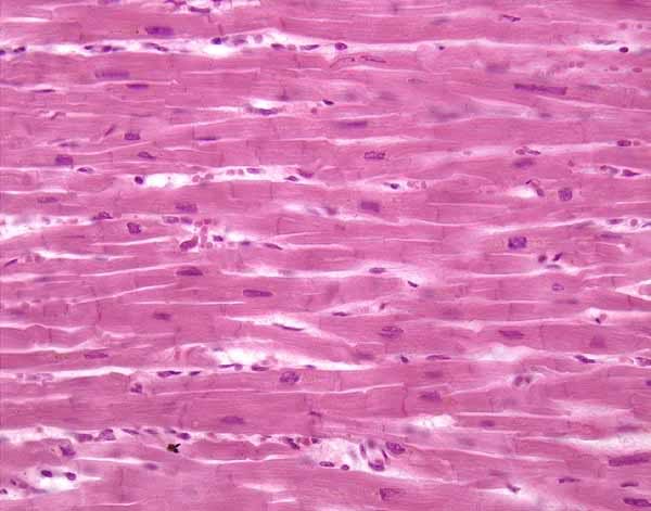
Cardiac Muscle Intercalated discs


More Cardiac Muscles

muscle cardiac intercalated

Connective Tissues Blood and Bone

Blood Features to know: Lymphocytes, Erythrocytes and Platelets

Lymphocytes


erythrocytes- blood cells


Bone Features to know:
Osteon
Oseteocytes
Lacunae
Canaliculi
Haversion Canal
Volkmann's Canal


Tuesday, April 8, 2008

Anatomy 302 Begins with Histology

There are various types of tissue.


Epithelial Tissue:

Simple Squamous- this photo shows Alveoli of the lungs


Stratified Squamous- Keratinized in skin


Pseudostratified Ciliated Columnar Epithelium


Simple Cuboidal Epithelium-


Simple Columnar Epithelium-


Transitional Epithelium-
Transitional epithelium

CT- Connective Tissue

Areolar CT


This image of areolar CT represents a whole mount (NOT a section) of mesentery (the tissue which binds together the loops of intestine within the peritoneal cavity.

This view looks through the entire mesentery, which is a sandwich of mesothelium on front and back surfaces with loose connective tissue and blood vessels in between.

The two most common cell types are mesothelial cells and fibroblasts.

The large, pale, oval nuclei belong to mesothelial cells which form the mesothelial surfaces on the front and back of the mesentery.

The smaller, darker, more elongate nuclei belong to fibroblasts, which manufacture and secrete the molecules which form the extracellular matrix.

Macrophages, mast cells, and lymphocytes may also be found in such preparations, but none are clearly identifiable in this image.

The matrix consists of unstained ground substance through which pass fibers of made of collagen and elastin.

Elastic fibers are thin, fairly uniform in diameter, and frequently branched. The are normally visible only if specially stained (as they have been in this specimen).

Collagen fibers vary in thickness and are eosinophilic (pink, in this stain).

CT- Elastic Fibers
CT Elastic tissue dermis

CT- Reticular CT silver stain only
CT Reticular fibers silver stain

CT- Hyaline Cartilage Chondrocytes in Lacunae
CT Hyaline cartilage

CT- Adipose fat storage
CT Adipose

Monday, March 10, 2008

Conduction System of the heart and Electrocardiography

Thanks for the photos Jesica!
smile thumbs up
Cardiac Chambers and Cycle


Cardiac Conduction System


1- Sinoatrial (SA) node- pacemaker
2- Atrioventricular (AV) node
3- AV bundle- Bundle of His
4- Bundle Branches
5- Perkinje Fibers


Cardiac Cycle


P Wave- Atrial depolarization
P-Q Interval- Atrial systole
QRS Complex- Ventricular depolarization begins after Apex
S-T segment- Ventricular Systole
Q-T Interval- Ventricular depolarization through repolarization

Tachycardia and Bradycardia


ECG/ EKG rhythm info


EKG/ECG Pictorial

Tuesday, March 4, 2008

Wednesday, February 27, 2008

Cat Arterial and Venous System, the 2 minute tutorial

Amusing YouTube Tutorial with a simple cat model with veins marked. Less grotesque, although the narrator is bit interesting to listen to.

Veins and Arteries- catastrophy

Yes, not savory... Thanks to the street feline who donated his body to science. What can you do? People, it's like Bob Barker says...

Anyways our pics for the week


Arterial System Shown Here
Red Pins:
1- Aortic Arch
2- Right Brachiocephalic Artery
3- Subclavian Artery
4- Common Carotid Artery
5- Axillary Arery
6- Brachial Artery

Venous System
White Pins/Yellow Pins:
1. Superior Vena Cava
2. Inferior Vena Cava
3. Azygos Vein
4. Brachiocephalic Vein
5. External Jugular Vein
6. Inter Jugular Vein
7. Subclavian Vein
8. Axillary Vein
9. Brachial Vein

Another Picture Same View


~~~~~~~~~~~~~~~~~~~

Lower body Venous and Arterial Pins


Arterial System Shown Here
Red Pins:
11. Renal Artery
12. Inferior Mesenteric Artery
13. Iliolumbar Artery
14. External Iliac Artery ** In humans there exist the Lt and Rt common Iliac Arteres
15. Internal Iliac Artery **On cats internal iliac arteries branch off med. Sacral Artery
16. Femoral Artery

Venous System
White Pins/Yellow Pins:
8.Axillary Vein
9.Brachial Vein
10. Renal vein
11. Gonadal vein
12. Common Iliac Vein
13. External Iliac Vein
14. Femoral Vein
15. Great Saphenous Vein


Another Picture same view:


2 More Views: 1 Without pins