Kidney terms to know:
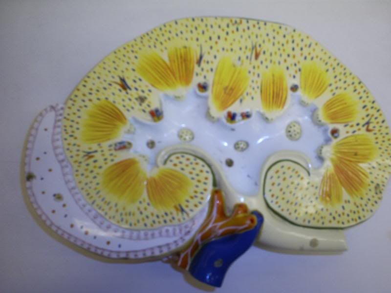
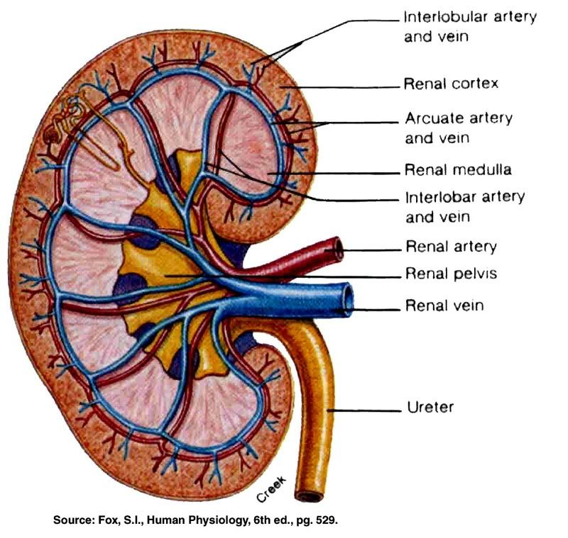
Hilium (hilius)- renal artery, vein and ureter enter/leave kidney
Renal Capsule- smooth transport membrane that adheres tightly to external aspect of kidney
Cortex- Superficial kidney layer- lighter in color and full of Bowman's capsules
Medulla- Deep to cortex, darker red/brown holds loops of henle
Medullary pyramids- triangular and striated sections, base facing cortex
papilla (apex)- points to inner kidney
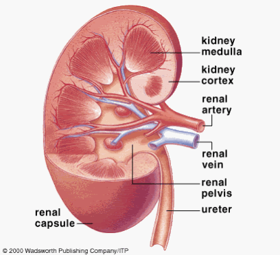
Renal Columns- area of cortex which dips between pyramids
Renal Pelvis- Medial to hilus, relatively flat/basin-like cavity w/ ureter
Major Calyx- large external of renal pelvis
Minor Calyx- subdivision of major calyx
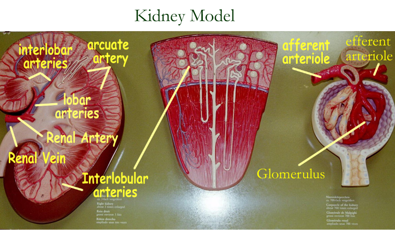
Renal Artery/Vein- branches 5x's as it enters the kidney it becomes --->
Segmental Arteries- enter hilus it becomes --->
Lobar Arteries- it becomes --->
Interlobar Arteries-
Aracate Arteries- at top of medullary region, these curve over base of medullary pyramid
Interlobular Arteries- ascend into cortex giving off--->
Afferent Arterioles
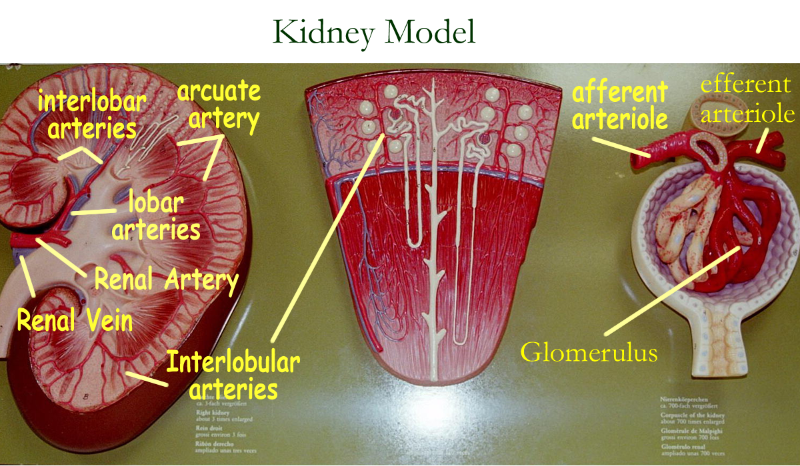
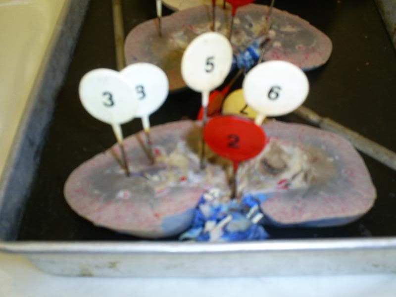
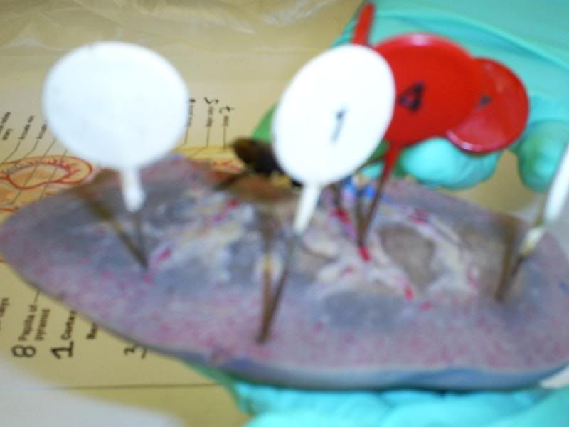
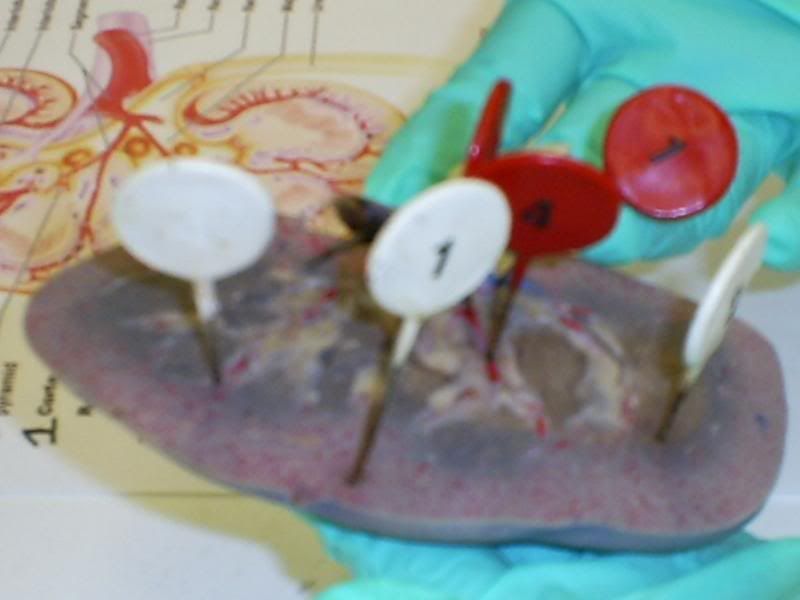
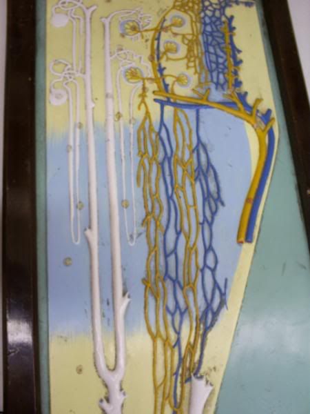
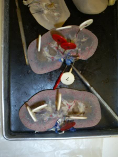
Pinned Models from class:
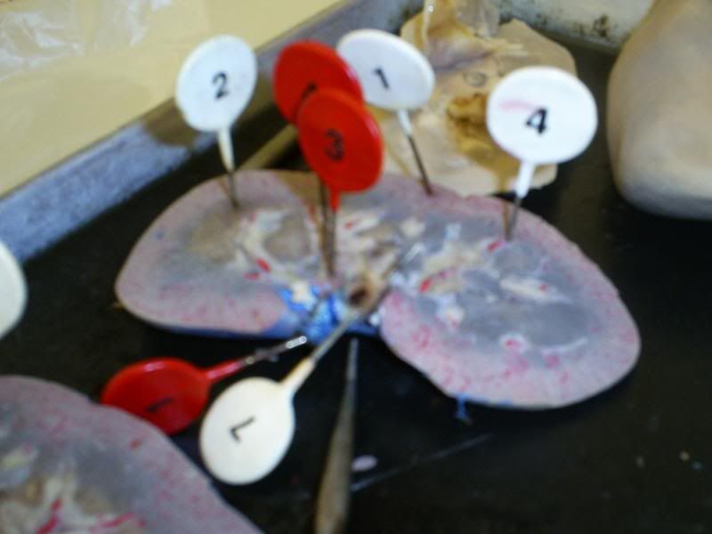
White-
1. Cortex
2. Renal Pyramid of Medulla
3. Minor Calyx
4. renal Column
5. Major Calyx
6. Renal Pelvis
7. Ureter
8. Papilla of Pyramid
Red
1. Renal Artery
2. Renal Vein
3. Segmental Arteries
4. Interlobar Artery
Tuesday, May 20, 2008
Histology of the kidney- A lot to process
Kidney terms to Know:
capsule
Cortex
medulla
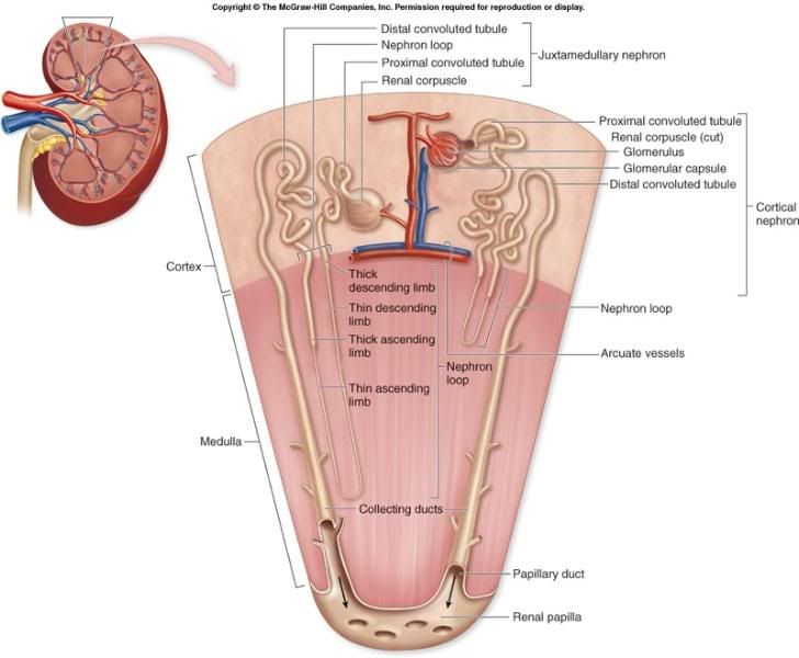
Cortical Nephron
Juxtamedullary Nephron
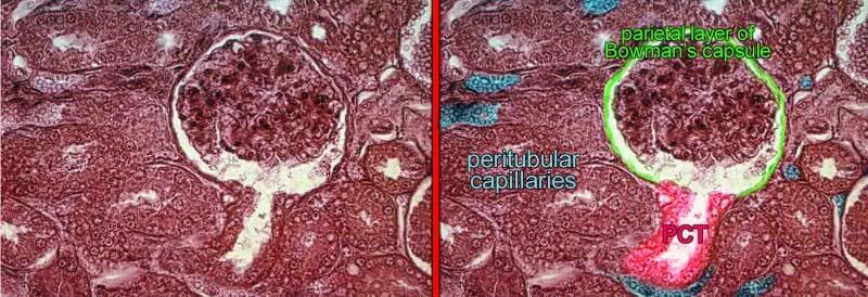
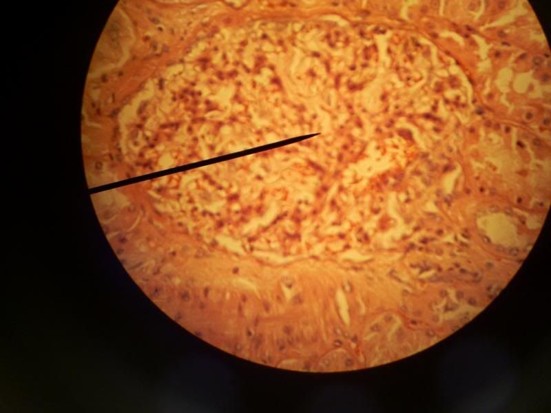
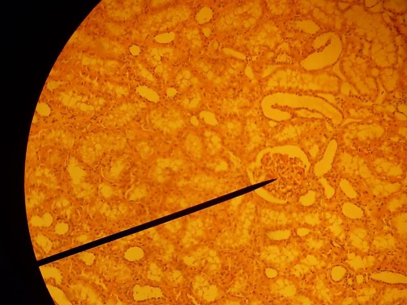
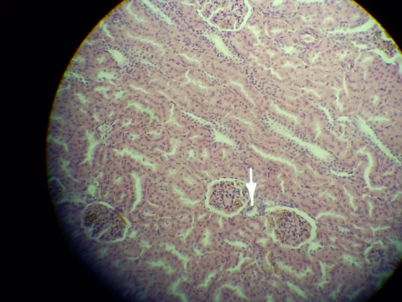
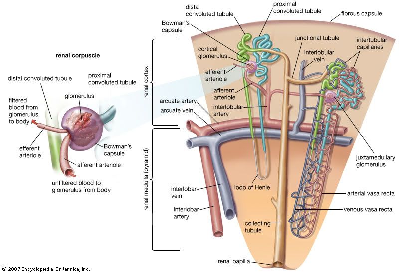
Renal Corpuscle
Glomerulus
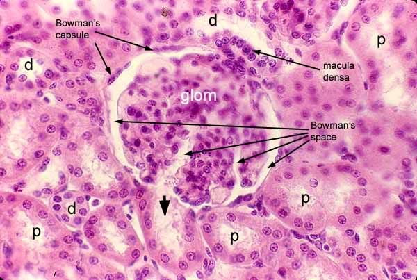
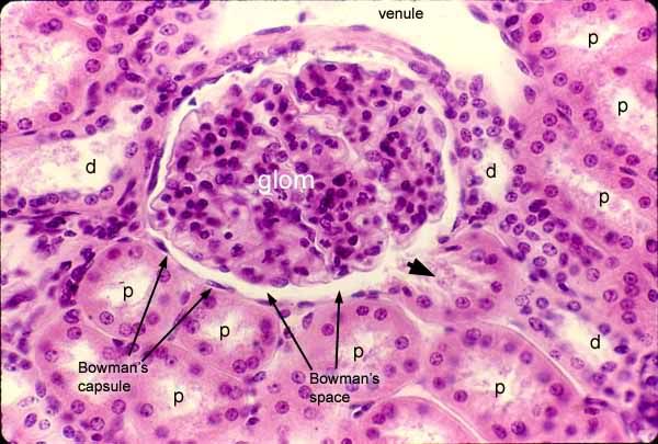
Bowman's Capsule (Glomerular Capsule)
visceral layer-consists of podocyte touching glomerulus
parietal layer- simple squamous
vascular pole- side w/ afferent arteriole and efferent arteriole & DCT
urinary pole- opposite of vascular pole
Bowman's Space
Podocytes
Proximal Convoluted tubule
Distal convoluted tubule
Macula densa
P are the PCT and D are the DCT
Loop of Henle
thick segment
thin segment
Collecting ducts
Papillary Ducts
Minor calyx
Medullary Ray
Kidney Lobule
Juxtaglomerular Cells
Juxtaglomerular Apparatus
Afferent Arteriole
Efferent Arteriole
vasa recta
Peritubular capillaries
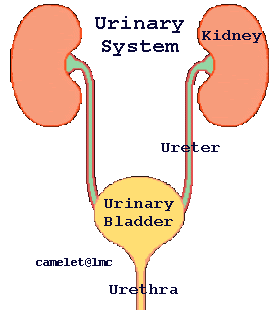
Bladder and Ureters histology that goes right through you
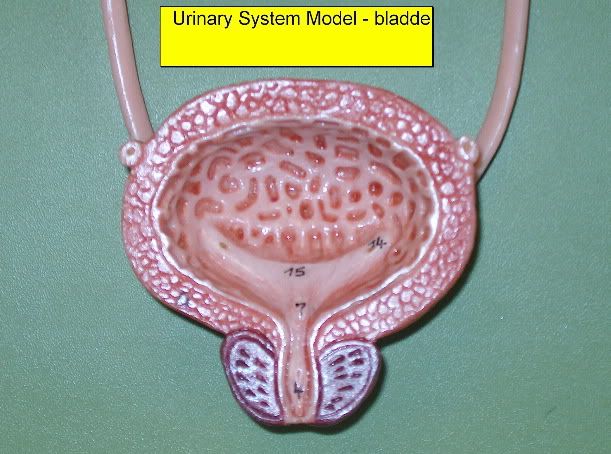
Bladder and Ureters terms to know:
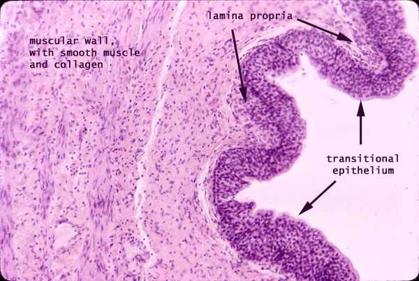
Transitional Epithelium
Subepithelial CT
lamina propria
submucosa
bladder and muscle layers
inner longitudinal
middle circular
outer longitudinal
Bladder muscle layers and transitional epithelium
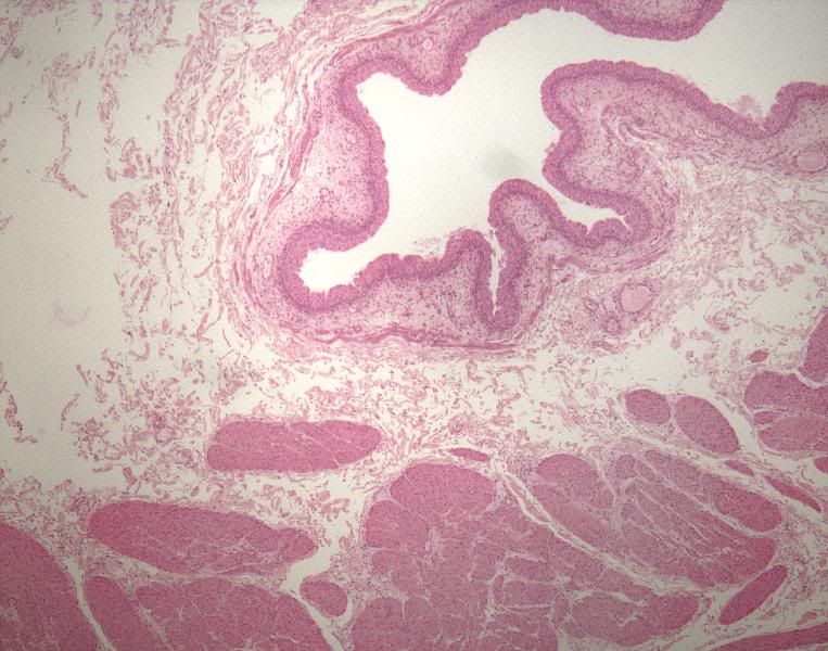
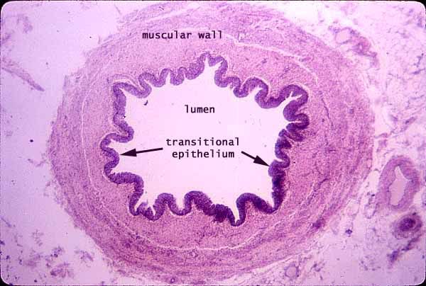
Stellate lumen of ureter- wavy epithelium
Ureter muscle layers
inner longitudinal
outer circular
Adventitia
Ureter muscle layers labeled
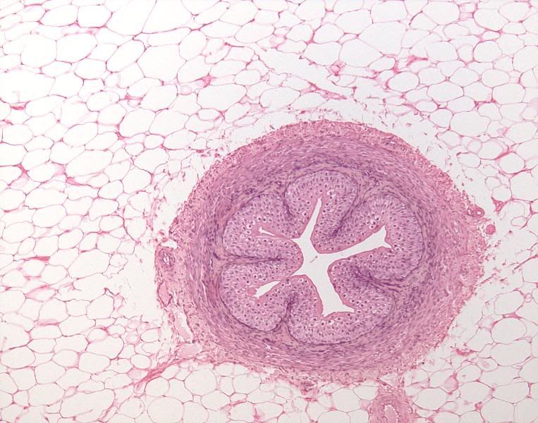
Unlabeled layers
Urethra muscle layers
a quick overview of bladder and ureter histology from Suny Downstate Medical Center... never heard of it, but the info is good.
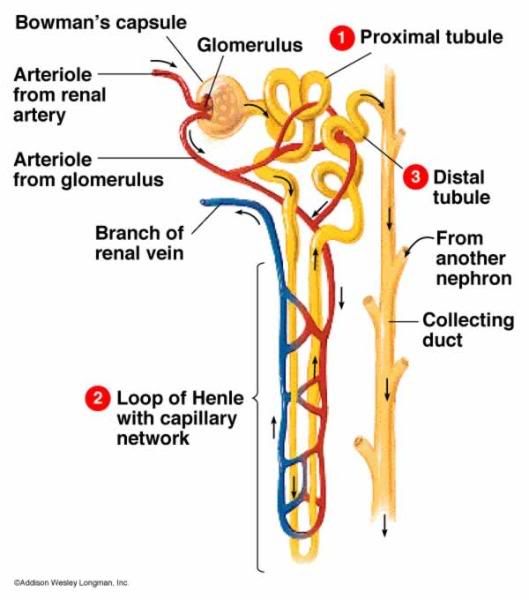
Pathways of fluid flow through kidney and urinary system
Great kidney info site by Vanderbilt University.. unfortunately the slides cannot be pulled up... only a few very good pics are accessible. vanderbilt U info
As usual, the best web site with a plethora of histo pics is SIU Med they pretty much rock the house of histo with tons of slides and great info as well...
Another good kidney histology site from some Tyler Junior College they have a few slides of each part of the kidney.
Histology from Pittsburg University The labels and info included are concise and easy to skim through fr review
Blood flow through kidney:
Renal Artery---> segmental artery---> lobar artery---> interlobar artery---> arculate artery---> interlobular artery---> afferent arteriole---> glomerular capillaries---> efferent arteriole---> peritubular capillaries---> vasa recta (juxtamedullary nephrons only) ---> interlobular vein---> arcuate vein---> interlobar vein--->renal vein
Blood flow through urine:
Glomerulus---> Bowman's capsule---> proximal tube---> descending loop of henle---> ascending loop of Henle---> DCT distal convaluted tubule---> collecting duct---> papillary duct---> minor calyx---> major calyx---> renal pelvis---> ureter---> bladder---> urethra---> and out of the body